About Corals, Anemones, and their Kin

Corals, anemones, and their relatives belong to the large and diverse phylum of animals called the Cnidaria.

General Information about Cnidaria

The basic body plan of members of this phylum is a simple sack with one opening that serves as both mouth and anus. In many species the mouth also functions as a genital orifice for the release of gametes or larvae. The fundamental sack-like structure is typically stretched out in certain areas giving the animal's body a more complex shape, most commmonly either a cylindrical body or a flattened cylindrical (or bell shaped) body, with a ring of tentacles surrounding the mouth. This simple radially symmetrical body consists of just two tissue types, ectoderm on the outside, and gastroderm on the inside, with varying amounts of jelly-like mesoglea between the two (amount varies among species); in the simplest cnidarians (e.g. freshwater hydra), the body consists of just two cell layers, a layer of epidermal cells lined internally by a layer of gastrodermal cells, though larger cnidarians are typically more complex.
 
There are two basic body forms among the cnidaria:
- polyps are typically sessile (attached in one place), though many anemones and some other polyps are capable of moving about slowly by various means.
 
- examples of cnidarians exhibiting the polyp body form include freshwater hydra, as well as anemones, corals, and others
 
- in many species, the polyps live in interconnected colonies (e.g., see discussion of stony coral structure below)
- this is typical of jellyfish and the dispersal stages of hydroids
 
- medusae are free-swimming, with swimming powered by contractions of the bell-shaped body
 
Polyp and medua in cross-section:


A medusa can be viewed as simply being a polyp flipped upside-down and stretched wide and flat:


A polyp


Upside-down polp


Upside-down polyp stretched - looks like a medusa (e.g. a jellyfish)


Many of the cnidarians alternate between the polyp body form and the medusa form.

For example, consider the life cycle of marine hydroids, which we typically think of as polyps (typically tree-like branching colonies of polyps. See photo at right of approx. 2cm long colony on wild-collected turtlegrass).
  1. Swimming, jellyfish-like medusae bud off from polyps.
  2. These medusae produce sperm and eggs.
  3. After fertilization, the eggs develop into ciliated, swimming planula larvae.
  4. The larvae settle out and attach on a suitable surface and develop into polyps.
  5. These polyps then bud additional polyps, which in colonial forms stay attached together to form colonies.


Feeding (and defense) and other modes of nutrition:
 
Feeding and defense:
 
Cnidarians have nematocysts, which are specialized structures (produced by certain cells) that function in food capture and defense. These nematocysts are pressurized and contain harpoon-like structures, complete with a "spear" that is connected to the animal by a "rope". These "harpoons" shoot out when the nematocyst is triggered by appropriate tactile and/or chemical stimuli. The fired nematocysts attach to or penetrate organisms that the cnidarian has come in contact with, often injecting poison in the process. These nematocysts not only aid in capturing animal prey or other food particles, but often they also serve to protect cnidarians from would-be predators or from other cnidarians competing for living space.
 
Cnidarians capture and eat a wide variety of prey. Depending on the cnidarian species, prey might include bacteria, protozoans, zooaplankton, fish, or other aquatic creatures. In addition, some cnidarians feed extensively on phytoplankton (free-living marine algae), some feed on small bits of organic debris (commonly referred to as "marine snow"), and some species absorb dissolved organic componds from the water.
 
Captured prey or other food particles are typically moved by the tentacles to the mouth and/or by cilia covering the body surface, and the food is taken into the gastrovascular cavity through the mouth. Digestion occurs within the gastrovascular cavity, and any undigestible portions of the food are later ejected through the mouth.
 
Other modes of nutrition - photosysnthesis by symbiotic algae:
 
Though most (or all?) cnidarians engage in some form of food capture as described above, many species also contain symbiotic dinoflagellate algae within their cells. These symbionts (called zooxanthellae) carry out photosynthesis and produce energy-rich food molecules, some of which leak out of the algal cells and into the surrounding cnidarian tissues. Many species of cnidarians depend on their algal symbionts for the majority of their energy needs, and loss of their zooxantheallae (as sometimes occurs under stressful conditions) can be lethal to the animal.
 
Cnidarians that depend on zooxanthellae include all of the reef-building stony corals, many of the soft corals and gorgonians, many species of anemomes, some jellyfish, and others.
 
(Note: in freshater habitats, green hydra also contain algal symbionts, but their symbionts are a species of green algae rather than dinoflagellates)


Cnidarian Diversity

There are three major groupings, or classes of cnidarians, which differ in the predominance of the two body forms in their life cycles:
Both among and within these three groups there is tremendous diversity of form and lifestyle, with cnidarians of one sort or another inhabiting most aquatic habitats. They are particularly abundant and diverse in marine (i.e. saltwater) environments, and in coral reefs, the reef structure itself is built largely by anthozoans in the order Scleractinia.
 
The overview of cnidarian diversity below focuses primarily on the Anthozoans, since this is the group best represented in Augsburg's aquaria.
 

(Based on similar diagram in: Delbeek, J. C, and Sprung, J., 1994. The Reef Aquarium, vol 1. Ricordea Publishing, USA.)


Overview of Stony Corals (Order Scleractinia)

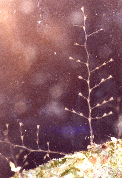
The importance of stony corals


Coral reefs are among the most species-rich habitats on earth, rivaled only by tropical rainforests in their diversity. Though the determinants of species diversity in ecosystems in general are not completely understood by ecologists, the high species diversity in coral reef ecosystems undoubtedly stems in part from the complex physical structure of these reefs, which provides diverse microhabitats for diverse life forms. This physical structure is created largely by the growth of stony corals, which build calcium carbonate skeletons as they grow. These coral skeletons (along with bits of shells and other hard parts of organisms), built up over thousands of years and cemented together by growths of coralline algae, make up the physical structure of coral reefs. At any given point in time, the currently living corals on the reef form a thin veneer of live tissue on the upper surfaces of the reef structure. The older skeletons below are riddled with crevices, caves and holes (bored out by sponges, worms, other organisms, and water currents) that are inhabited by diverse forms of life.
 

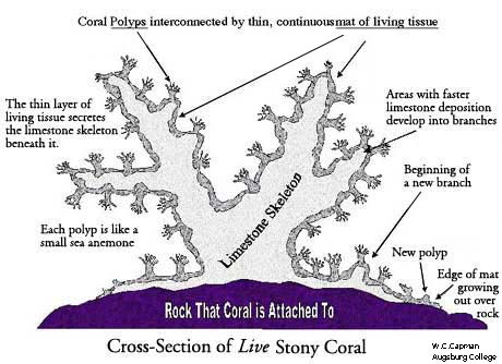
Stony coral structure and colony development

 
At first glance, it can be difficult to relate the structure of a stony coral to the generalized polyp body plan. What you typically notice first about a stony coral is not the structure of the polyp itself, but rather the structure of the colony, which can often consist of many thousands of polyps. Furthermore, in many species the polyps are partially or completely retracted during the day or are extremely small. In other cases (e.g. brain corals ond others), the basic polyp body plan is so modified that it can be a challenge at first to see any relation between what you see and polyp structure (more on this follows diagram below).
 
To understand the structure of a stony coral colony, it is worthwhile to consider how a coral colony develops:
  1. A stony coral colony generally starts out life as a swimming, planktonic, ciliated larva called a planua. . Most planulae either get eaten by predators or are taken by currents to unsuitable locations, but some ultimately find suitable hard surfaces where they settle and attach.

    (Note: in some cases the planula stage is bypassed. In many corals, fragments that break off colonies can attach to rocks and develop into new colonies (which is how most captive propagation of corals is accomplished), or individual polyps or small balls of tissue spontaneously released from large colonies can attach and found new colonies).

  2. After settling, the planula develops into a polyp. This polyp grows and starts to secrete a calcium carbonate (limestone) skeleton beneath it.

  3. In colonial species, the polyp soon starts to bud off new polyps from the base, which generally remain attached to the original polyp and to each other by a continuous mat of living tissue. Skeleton deposition continues beneath this mat.

    - In many species, this mat of tissue originating from the base of the founding polyp grows out first, encrusting the substrate, and the new polyps develop from this mat (see diagram below).

    - In other cases new polyps don't bud from the base, but rather large polyps elongate (so the ring of tentacles becomes oval shaped), the elongated polyp narrows and pinches off in the middle, and the polyp is split in two. The boulder coral in Augsburg's reef aquarium produces new polyps this way.

  4. If the colony secretes calcium carbonate skeleton fairly uniformly underneath this mat of tissue, the colony develops into a mound, ball, or boulder shape. Alternatively, if some areas grow and secrete calcium carbonate faster than others, the faster-growing regions develop into branches. The result of the latter form of growth is diagrammed below.


While the individual polyps in corals such as the one diagrammed above actually look like polyps, in many corals the basic polyp shape has been highly modified. A few examples:
 
Small vs. Large Polyped Stony Corals (SPS vs. LPS corals):
 
Polyp size varies greatly among stony corals, ranging from some with polyps around a millimeter or two in diameter to others with polyps that can exceed diameters of 30 cm (1 foot). Stony corals with very small polyps are often referred to as small-polyped scleractinian corals (or SPS corals for short), while larger polyped stony corals are called large-polyped scleractinian corals (or LPS corals for short). Though there is no clear demarcation between these two informal categories, those referred to as LPS corals generally have polyps ranging from a centimeter or so in diameter up to the largest sizes, while polyps of SPS corals tend to be much smaller.


Things to look for in future updates of this page